"Third Eye" Spinal Removal Treatment
- incisionary
- Dec 26, 2025
- 3 min read


In May of 2025, a grand surgical innovation was made with the creation of a procedure designed to remove a tumor at the junction of the skull base & upper spinal cord. More colloquially noted as a “third eye” tumor removal procedure, a transorbital endoscopic spinal tumor removal is a new neurosurgical procedure that allows for a tumor between the skull base and a portion of the spinal cord to be removed. With this procedure, surgeons hoped to remove tissue creating pressure on the spinal cord without doing a larger surgery that would likely require some bone removal or chances of compromising the person’s spine as a whole. People have historically approached spinal procedures by going directly through the spine to operate. This procedure, however, allows for access to the spine through one’s natural anatomy. Surgeons involved in the first attempts at this procedure went through the eye socket to get to the spine.
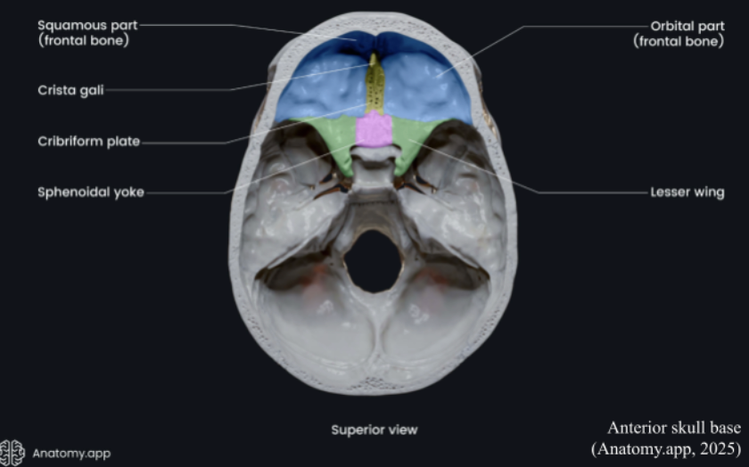
Extensive detailed imaging and surgical planning were required to map out the tunnel the surgeons would take to get to the person’s spine. Images of the person’s blood vessels, nerves, and bones were created to allow for such planning. After the patient was prepped for surgery, the surgeons began creating their tunnel to the spinal cord by only removing a small section of the bone with precision drills designed for accuracy and careful cuts. They did this to get access to the anterior skull base without potentially disturbing the person’s brain. Endoscopic technology is leaned on heavily throughout this procedure because it allows for high-definition, high-quality imaging. An endoscope is inserted through the nasal cavity to allow for a fully dimensional viewing of the body’s internal structures. With this, surgeons are able to make more accurate movements. Surgical instruments are inserted through the small opening made prior to the insertion of the endoscope, which helps guide surgeons as they move their instruments towards the tumor for removal. Once they’re able to reach the tumor, surgeons begin separating it from the spinal cord, cranial nerves, and surrounding blood vessels. They take their time to remove the tumor by removing it in small sections, rather than approaching the entire tumor at once like in other tumor removal procedures. Microsurgical tools (extremely fine tools used with microscopes during surgery for miniscule procedures) are used to carefully dissect and extract the tumor tissue layers.
The lentitude of the removal also helps to preserve healthy structures and make sure nothing is removed that doesn’t need to be, seeing as the spine is an extremely delicate structure. Bleeding is controlled using other fine surgical tools that create pressure, making it so no new blood enters and no other blood leaves. Following the completion of dissection of the spinal tumor, intense inspection is done on the area to assure no parts of the tumor have been left behind. The area of surgery is closed with suturing, and all structures are put back into their regular positions. The cleanup process is just as delicate and careful as the process of getting to the area of the tumor. Surgeons must be extremely careful throughout their actions during this procedure because of how delicate the spinal cord is.
Written by Kamila Dessus at Incisionary
References
2025 News - In First-of-Its-Kind Surgery, Rare Spinal Tumor Removed Through Patient’s Eye Socket at University of Maryland Medical Center | University of Maryland School of Medicine. (2025). Umaryland.edu. https://www.medschool.umaryland.edu/news/2025/in-first-of-its-kind-surgery-rare-spinal-tumor-removed-through-patients-eye-socket--at-university-of-maryland-medical-center.html
Anatomy.app. (2025). Anterior cranial fossa | Encyclopedia. Anatomy.app. https://anatomy.app/encyclopedia/anterior-cranial-fossa
Clinic, C. (2024). Microsurgery: What It Is, Procedures & Instruments Used. Cleveland Clinic. https://my.clevelandclinic.org/health/treatments/microsurgery
First-of-Its-Kind Surgery Uses Eye Socket to Remove Spinal Cancer. (2025, May 15). Ophthalmology Advisor. https://www.ophthalmologyadvisor.com/news/first-of-its-kind-surgery-uses-eye-socket-to-remove-spinal-cancer/
Graham, F. (2025). Daily briefing: A spinal tumour was removed through a person’s eye socket for the first time. Nature. https://doi.org/10.1038/d41586-025-01441-0
Johnson, M. (2025, May 4). Surgeons bid for medical first: Removing spinal tumor through patient’s eye. The Washington Post.
Julia. (2025, June 2). Rare spinal tumour removed through eye socket. Surgery International. https://surgery.international/rare-spinal-tumour-removed-through-eye-socket/
UMMCVideos. (2025, June 3). Your Health: First of its kind spinal tumor surgery. YouTube. https://www.youtube.com/watch?v=EK9g1pxHuGU


Comments